
GDV (bloat) in dogs: the golden one hour
Of all the emergencies I see, gastric dilatation-volvulus is the one where the clock is most visible. In a dog with a simple foreign body, you have hours. In a dog whose stomach has twisted, you have one hour to get him on the table if you want the best odds. After two hours the mortality curve bends sharply upward. After four, we are often talking about damage that cannot be undone.
This article is the timeline I want every owner of an at-risk dog to have memorized before they ever need it.
What GDV actually is
GDV is two problems stacked on top of each other. First the stomach fills with gas, fluid, and foam — that's the dilatation. Then, usually because the dog is active or rolls, the distended stomach rotates on its mesenteric axis, twisting somewhere between 90 and 360 degrees. Once twisted, nothing moves in or out. Gas keeps fermenting, the stomach balloons against the diaphragm, and the splenic vessels kink off.
Three things kill the dog in the next minutes-to-hours:
- Cardiovascular collapse. The dilated stomach compresses the caudal vena cava. Venous return to the heart drops, blood pressure plummets, and the dog goes into obstructive and hypovolemic shock simultaneously. Heart rates above 180 bpm and pale or muddy gums are the rule, not the exception.
- Gastric wall necrosis. Twisted vessels cut off perfusion. After 4–6 hours, portions of the stomach wall are dead. Necrotic tissue is removed surgically; the dog that needs more than a partial gastrectomy is the one whose odds fall off a cliff.
- Reperfusion injury and arrhythmia. Even after successful surgery, the sudden return of blood flow to ischemic tissue floods the circulation with potassium and acid byproducts. Ventricular arrhythmias appear in roughly 40% of post-op GDV patients, usually within 12–24 hours. They are the single biggest killer of dogs who survived the first surgery.
Who is at risk
GDV is overwhelmingly a disease of large, deep-chested breeds. The classic list:
- Great Dane (lifetime risk roughly 1 in 4 — the highest of any breed)
- Standard Poodle, Weimaraner, Saint Bernard, Irish Wolfhound
- German Shepherd, Boxer, Doberman, Gordon Setter
- Basset Hound and other deep-but-low chested breeds
- Any mixed-breed dog with a narrow chest-to-depth ratio
Risk climbs with age — most cases are in dogs 7 years and older — and with several modifiable factors: eating one large meal a day, eating very fast, eating from a raised bowl, exercising within an hour of eating, and a nervous or fearful temperament. Having a first-degree relative that bloated roughly doubles the risk, suggesting a heritable chest shape.
If you own a dog in this list, you should be thinking about prevention before you are thinking about symptoms.
The signs you will see in the first hour
GDV does not start subtle. The textbook progression in the first 30–60 minutes is:
- Non-productive retching. The dog stands with his head down, mouth open, gagging — nothing comes up, or only a thin rope of white foam. This is the single most specific sign and the one I ask about first.
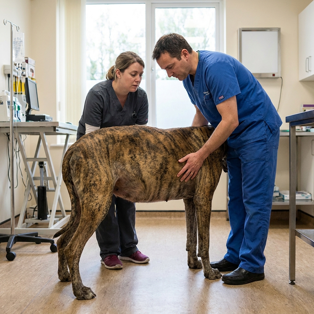
- Abdominal distension. The left flank behind the rib cage looks visibly swollen and feels tympanic (drum-tight) when you tap it. In a heavily muscled or long-haired dog, look from above and compare both sides.
- Drooling and restlessness. Salivation increases, the dog cannot settle, paces, looks at his flanks.
- Rapid deterioration. Within an hour, gums turn pale or muddy, heart rate climbs above 160 bpm, and the dog becomes weak or collapses.
A quick home screen I teach owners: if a large deep-chested dog is retching without producing vomit and his belly is getting visibly larger while you watch, that is the drive-now combination. Do not wait for collapse. Do not offer food. Do not "see how he is in an hour."
What happens at the clinic: the first 60 minutes
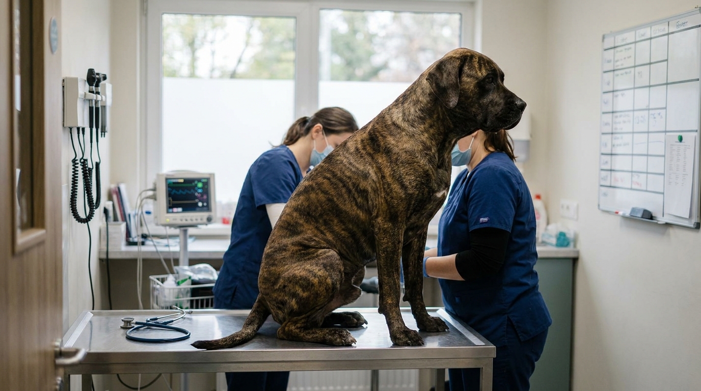
When you arrive, the team has about 10 minutes to convert "GDV or not" into "on the table or not." The protocol I run is the same one I would want run on my own dog.
1. Confirm in one X-ray. A right lateral abdominal radiograph is the standard view. The dilated, gas-filled stomach sitting in the cranial abdomen looks like a "double bubble" or, classically, a "Smurf hat" — a sharply divided compartment with a soft-tissue line down the middle where the pylorus has flipped over the fundus. A simple bloat (dilatation without volvulus) shows a single gas bubble; volvulus shows two.
2. Decompress immediately. A percutaneous trocar (large-bore 14–16 gauge needle) is passed through the skin into the stomach, releasing gas with a characteristic hiss. This is fast and buys minutes. After trocarization, an orogastric tube is passed through the mouth to fully empty the stomach — this requires light sedation because the tube is large and the dog is awake.
3. Shock-dose IV fluids. Two large-bore catheters go in. Crystalloids — lactated Ringer's or Plasma-Lyte at 60–90 mL/kg in the first hour — are given as rapid boluses. Many of my colleagues add hypertonic saline 7.5% at 4–5 mL/kg over 5–10 minutes to pull fluid out of the swollen splanchnic bed and into the circulation, but only in the first minutes and only if the dog's sodium is not already high.
4. Analgesia, carefully. The dog is in severe pain but is also in shock. Pure mu-opioids are safe in shock: hydromorphone 0.05–0.1 mg/kg IV, methadone 0.3–0.5 mg/kg IV, or fentanyl 3–5 µg/kg IV as a bolus. I avoid acepromazine and the alpha-2 agonists (medetomidine, dexmedetomidine) in shock patients because both drop blood pressure further. I also avoid NSAIDs until perfusion and renal function are stable — usually 24–48 hours post-op.
5. ECG monitoring from the start. A 12-lead or at least continuous lead-II ECG runs the entire stabilization. Ventricular premature contractions are treated with lidocaine 2 mg/kg IV bolus (slow push), then a CRI at 25–80 µg/kg/min if the arrhythmia persists. Lidocaine doses in dogs are real — a 25 kg dog gets 50 mg bolus, then a 12.5–40 mg/hour drip.
6. Antibiotic cover. Gastric wall compromise lets bacteria translocate. A first-generation cephalosporin such as cefazolin at 22 mg/kg IV at induction, repeated every 90 minutes during surgery, is my standard.
The dog that stabilizes goes straight to surgery. The dog that does not stabilize first rarely survives surgery, no matter how good the surgeon is.
The surgery itself
The goals, in order, are: derotate the stomach, assess the spleen and stomach wall, resect any dead tissue, and prevent recurrence with a gastropexy.
Derotation is counter-clockwise when viewed from behind the dog — the surgeon's first job is to identify the correct direction. The spleen is examined; if it is twisted and necrotic, it is removed (splenectomy), which the dog tolerates well. A gastric resection is performed only where the wall is dead; a partial gastrectomy is a guarded-prognosis procedure.
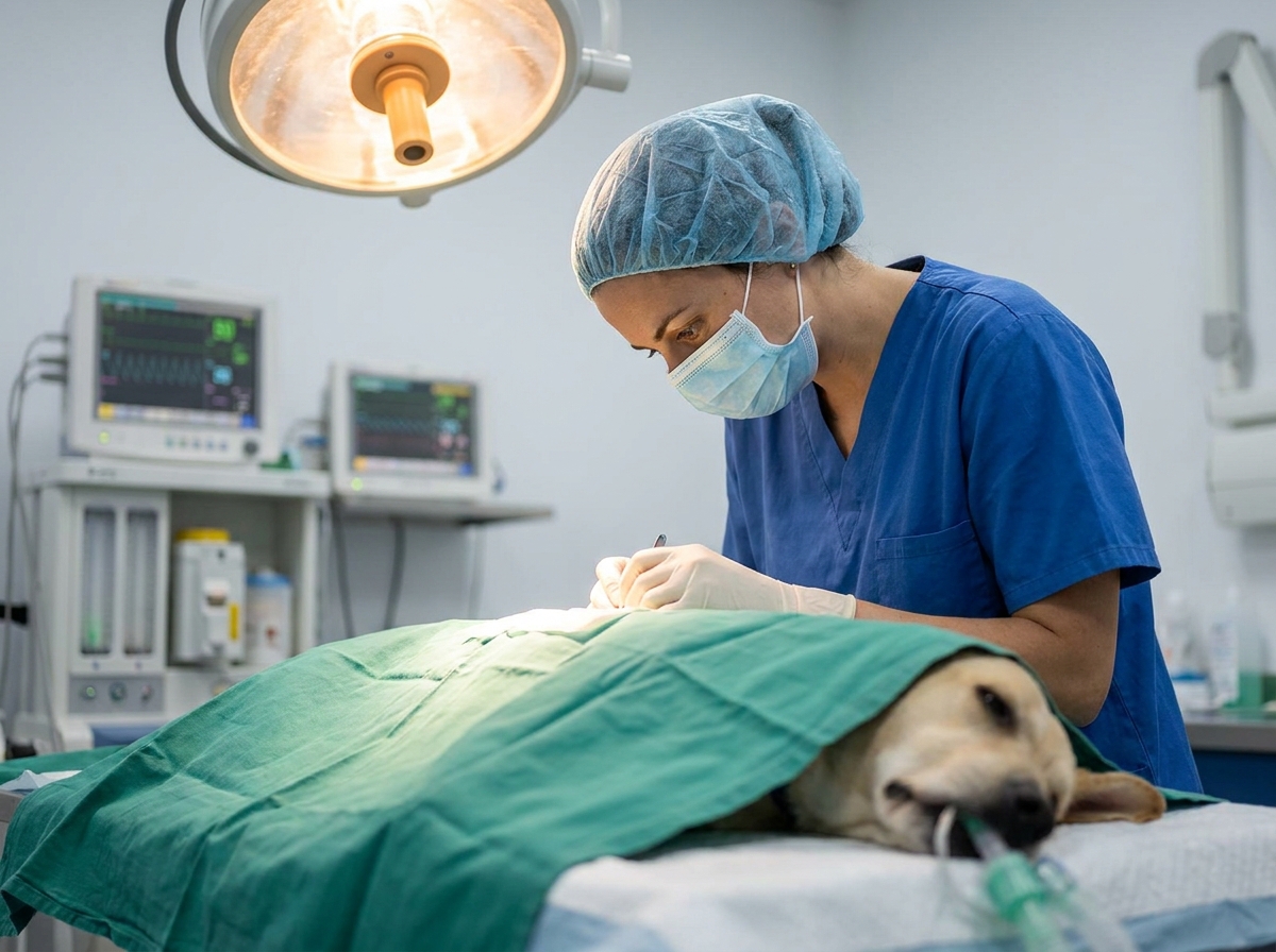
The procedure that prevents the next episode is a gastropexy — suturing the stomach wall to the right body wall so it cannot twist again. Studies show recurrence rates drop from over 80% in un-pexied dogs to under 5% in dogs that have a proper gastropexy. There is no good reason to leave this out.
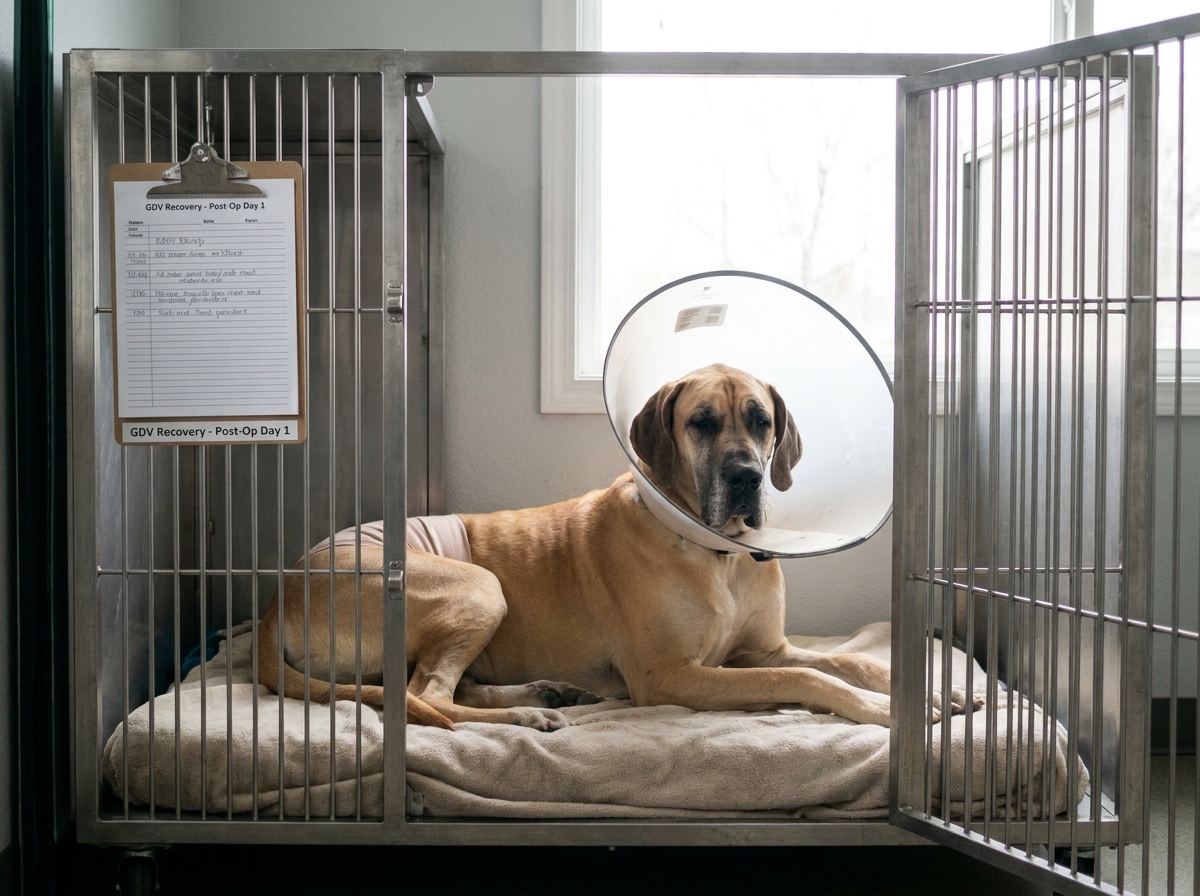
The 72 hours after surgery
This is the second cliff. Most dogs that die post-op do so in the first three days from one of three things:
- Reperfusion arrhythmia. Continuous ECG for at least 24–48 hours. Treat with lidocaine as above. Magnesium supplementation (MgSO4 0.2 mEq/kg IV over 15 min) is a useful adjunct when arrhythmias persist despite lidocaine.
- Sepsis or peritonitis from a gastric resection or a missed perforation. Watch for fever, abdominal pain, falling albumin, and rising lactate.
- Aspiration pneumonia from the orogastric tube. Elevated head, suction available, and antibiotics if it develops.
Most dogs who sail through 72 hours go home in 3–5 days and live normal lives.
Prevention: what actually lowers risk
For a high-risk dog, I recommend three changes before anything goes wrong:
- Two or three meals a day, not one. Slow the eating with a slow-feeder bowl or by spreading kibble on a baking sheet.
- No vigorous exercise for 60–90 minutes before and after meals.
- Prophylactic gastropexy. Done laparoscopically or as an open procedure at the time of spay/neuter in a young Great Dane, Standard Poodle, or similar breed. It converts a future 30%-mortality emergency into a surgery the dog has already had. In breeds with a 1-in-4 lifetime risk, the cost-benefit math is straightforward.
If you have an at-risk dog, save the number of your nearest 24-hour emergency clinic in your phone tonight. And remember the single best diagnostic question: is he retching without producing vomit? If yes, you are on the clock.
---
This article reflects general emergency practice patterns; exact drug doses and protocols vary between hospitals and are tailored to the individual patient. It is informational and does not replace an in-person exam. If you suspect GDV, head directly to the nearest emergency clinic.