
Feline eosinophilic granuloma complex: the three presentations, the diagnosis, and the treatment
Feline eosinophilic granuloma complex (EGC) is a group of three related skin conditions in cats, all of which are driven by an overactive immune response involving eosinophils (a type of white blood cell). The three presentations are the indolent ulcer, the eosinophilic plaque, and the linear (or collagenolytic) granuloma. The three presentations can occur separately or together, and the three presentations can be present in the same cat at different times. The cause is most often an underlying allergic disease — flea allergy, food allergy, or environmental allergy (atopic dermatitis). The diagnosis is by skin cytology and skin biopsy, and the treatment is by addressing the underlying allergy plus, in many cases, anti-inflammatory medication. In this article I will walk through each of the three presentations, the diagnostic workup, the treatment, the prognosis, and the most common mistakes in management.
The indolent ulcer (rodent ulcer)
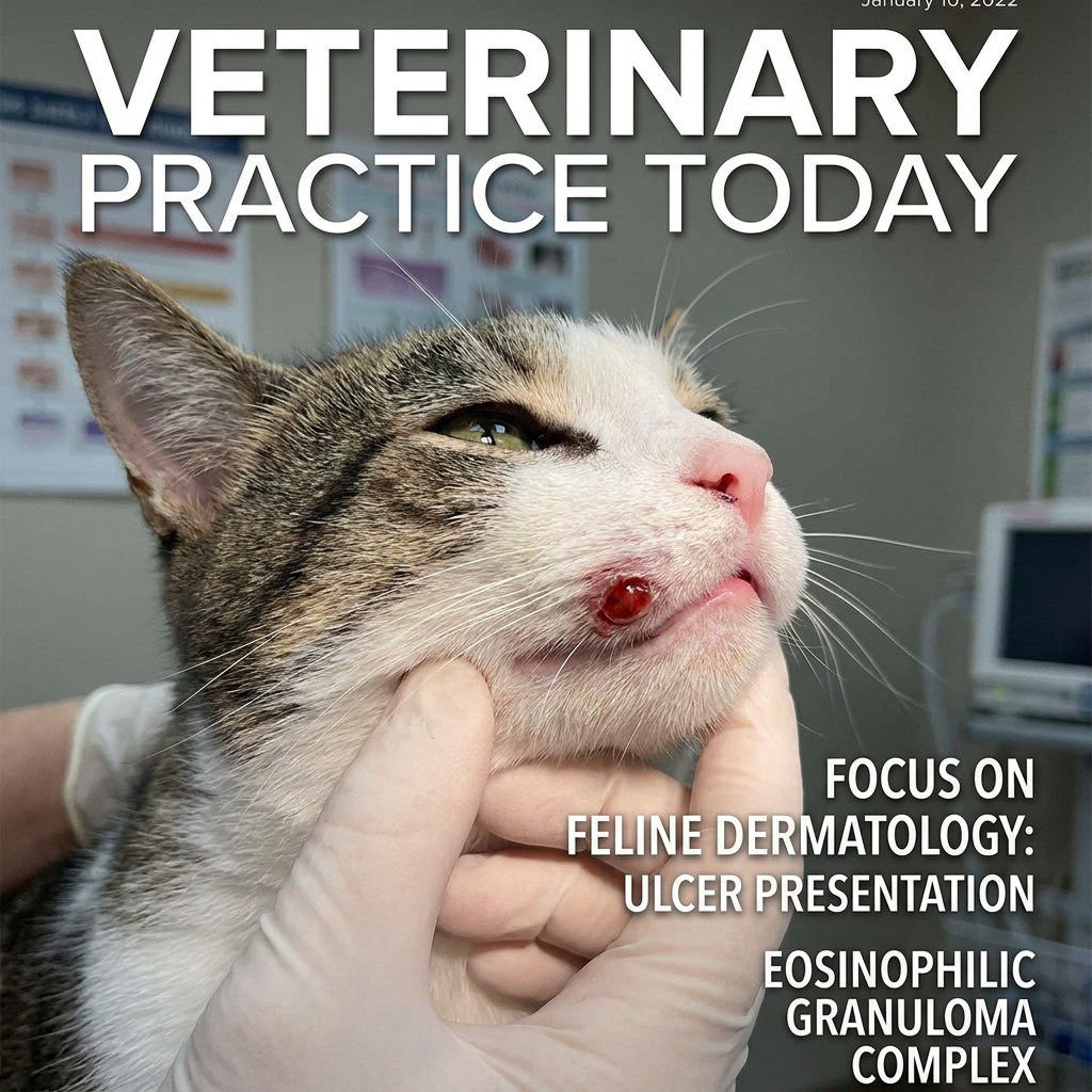
The indolent ulcer, also called the "rodent ulcer" because of the historical but incorrect belief that the ulcer was caused by rodents, is the most common presentation of EGC. The indolent ulcer is a well-defined, red-brown, glistening ulcer on the upper lip, near the philtrum. The ulcer is typically 2-10 mm in diameter, and the ulcer is often unilateral (on one side) or bilateral (on both sides). The ulcer does not bleed, does not appear to be painful, and does not typically affect the cat's appetite or behavior.
The cause: most often flea allergy, but can also be food allergy or environmental allergy. The cause can also be idiopathic (no identifiable underlying allergy). The diagnosis is by visual inspection (the appearance is characteristic), supplemented by skin cytology to confirm the eosinophilic inflammation.
The treatment: addressing the underlying allergy (flea control, food trial, environmental control) plus topical or systemic anti-inflammatory medication. The topical medication is often a potent steroid cream applied to the ulcer. The systemic medication is often a short course of oral prednisone or methylprednisolone. The ulcer typically responds within 2-3 weeks.
The eosinophilic plaque
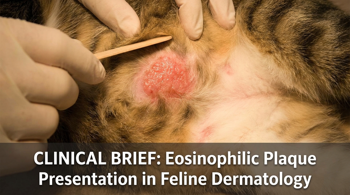
The eosinophilic plaque is the most pruritic (itchy) of the three presentations. The plaque is a well-defined, raised, red, hairless, moist, eroded lesion, typically 1-5 cm in diameter. The plaque is most commonly found on the ventral abdomen, the inner thigh, the neck, or the shoulder. The plaque is intensely itchy, and the cat that has the plaque is the cat that is scratching, licking, or biting the plaque constantly.
The cause: most often flea allergy or environmental allergy. The plaque is the most allergic-driven of the three presentations, and the plaque is often the first sign of a new allergy or a flare of an existing allergy.
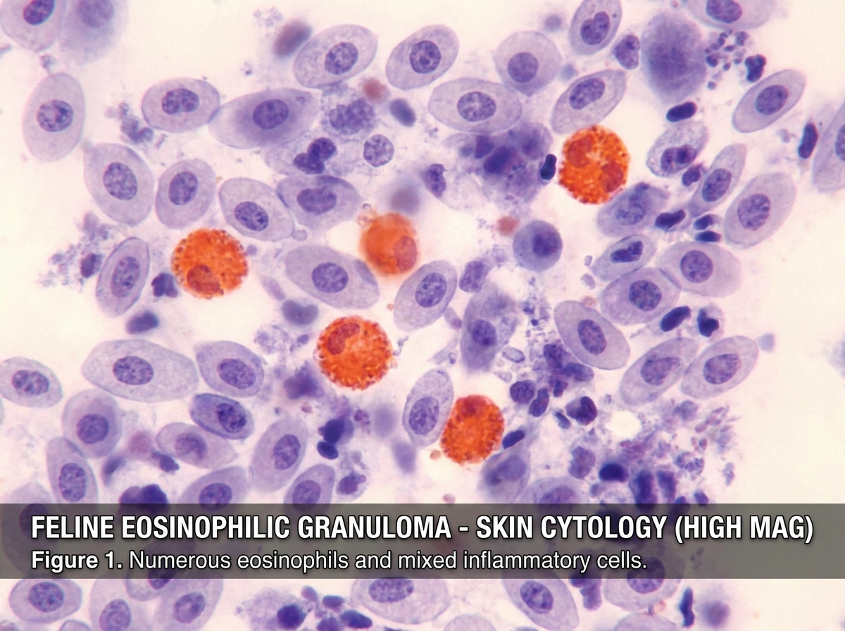
The diagnosis: skin cytology shows a large number of eosinophils, often with secondary bacterial or yeast infection. The skin biopsy is diagnostic if the cytology is inconclusive. The diagnostic workup for the underlying allergy: a flea comb, a food trial, an environmental allergy panel (intradermal or serum testing).
The treatment: addressing the underlying allergy plus anti-inflammatory medication. The most effective systemic medication is glucocorticoids (prednisone or methylprednisolone) at anti-inflammatory doses, or cyclosporine (Atopica) for the long-term management. The antibiotic or antifungal is added if the secondary infection is present. The plaque typically responds within 2-4 weeks, but the plaque often recurs if the underlying allergy is not controlled.
The linear (collagenolytic) granuloma
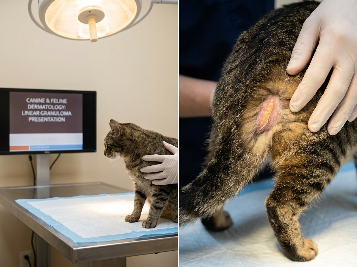
The linear granuloma is the least common of the three presentations, but the linear granuloma is the most distinctive. The linear granuloma is a firm, raised, yellow-pink, hairless, linear or oval lesion, typically 2-10 cm in length. The linear granuloma is most commonly found on the caudal thigh, the chin, or the oral cavity (the tongue or the palate). The linear granuloma in the oral cavity is the "oral linear granuloma," and the oral form can cause difficulty eating or drooling.
The cause: most often environmental allergy, but can also be flea allergy. The linear granuloma is the most steroid-responsive of the three presentations, often responding to a single course of glucocorticoids.
The diagnosis: skin cytology and skin biopsy, with the biopsy showing the characteristic "collagenolytic" (collagen-dissolving) inflammation. The diagnostic workup for the underlying allergy is the same as for the plaque.
The treatment: glucocorticoids (prednisone or methylprednisolone) at anti-inflammatory doses, with a rapid response expected. The oral linear granuloma may require a longer course of treatment, and the refractory oral linear granuloma may need additional immunosuppressive medication (chlorambucil or cyclosporine).
The diagnostic workup
The workup for a cat with suspected EGC: a full physical exam, a skin cytology (looking for the eosinophils), a skin biopsy if the cytology is inconclusive, a flea comb to look for fleas or flea dirt, a food trial (8-12 weeks on a novel or hydrolyzed protein diet) to rule out food allergy, and an environmental allergy workup (intradermal skin testing or serum IgE testing) to identify the specific environmental allergens.
The order of the workup: flea control first (the most common cause, and the easiest to rule out), then food trial (the second most common cause, and the second easiest to rule out), then environmental allergy workup (the third most common cause, and the most complex to work up). The cat that has all three workups negative is a cat with idiopathic EGC, and the treatment is symptomatic control with anti-inflammatory medication.
The treatment principles
The treatment of EGC has three layers: the underlying allergy, the secondary infection, and the anti-inflammatory medication.
The underlying allergy: flea control (a monthly topical or oral adulticide plus environmental control), food trial (8-12 weeks on a novel or hydrolyzed protein diet with no other food sources), environmental control (air purifier, frequent cleaning, avoidance of identified allergens). The underlying allergy is the cause of the EGC, and the underlying allergy is what must be controlled for the long-term resolution.
The secondary infection: bacterial (a course of antibiotics for 2-4 weeks), yeast (a topical or systemic antifungal). The secondary infection is what makes the lesion more inflamed, and the secondary infection is what slows the healing.
The anti-inflammatory medication: glucocorticoids (prednisone, methylprednisolone, or triamcinolone) for the short-term control, cyclosporine for the long-term control. The glucocorticoids are fast-acting and effective, but the glucocorticoids have side effects with long-term use. The cyclosporine is slower-acting (4-8 weeks for the full effect) but has fewer side effects with long-term use. The choice depends on the severity of the EGC, the response to the initial treatment, and the cat's ability to tolerate the side effects.
The prognosis
The prognosis for EGC is good with treatment. The cat that has the underlying allergy identified and controlled is the cat that has the long-term resolution. The cat that has the idiopathic EGC is the cat that requires long-term medication, with the medication adjusted as the disease waxes and wanes. The cat that is left untreated is the cat that has chronic, progressive skin lesions, secondary infections, and a significant impact on the quality of life.
The recurrence is common. The EGC that resolves with the initial treatment is the EGC that often recurs within 3-12 months, particularly during the allergy season. The owner who is prepared for the recurrence, who has the medication on hand, and who starts the medication at the first sign of the recurrence is the owner who manages the EGC effectively.
The most common mistakes
The first mistake: ignoring the underlying allergy. The owner who treats the EGC with steroids without identifying the underlying allergy is the owner who has the EGC recur every 3-6 months. The fix: the workup for the underlying allergy is essential, even if the workup is expensive or time-consuming.
The second mistake: stopping the medication too early. The owner who stops the medication when the lesion has resolved is the owner who has the lesion recur within 2-4 weeks. The fix: the medication is continued for 2-4 weeks after the lesion has resolved, and the medication is then tapered gradually over 4-8 weeks. The gradual taper is what allows the immune system to maintain the resolution.
The third mistake: not treating the secondary infection. The owner who treats the EGC with steroids alone, without addressing the secondary bacterial or yeast infection, is the owner who has a slow or partial response. The fix: antibiotics or antifungals as indicated, often in combination with the steroids.
The one-page summary
Feline eosinophilic granuloma complex has three presentations: the indolent ulcer, the eosinophilic plaque, and the linear granuloma. The cause is most often an underlying allergy (flea, food, or environmental). The workup is for the underlying allergy, plus the cytology or biopsy to confirm. The treatment is the underlying allergy control, the secondary infection treatment, and the anti-inflammatory medication. The prognosis is good with treatment, and the recurrence is common. The owner who runs the workup, who addresses the underlying allergy, who treats the secondary infection, and who continues the medication through the taper is the owner whose cat has the long-term resolution. The workup is the work. The workup is what makes the difference.