

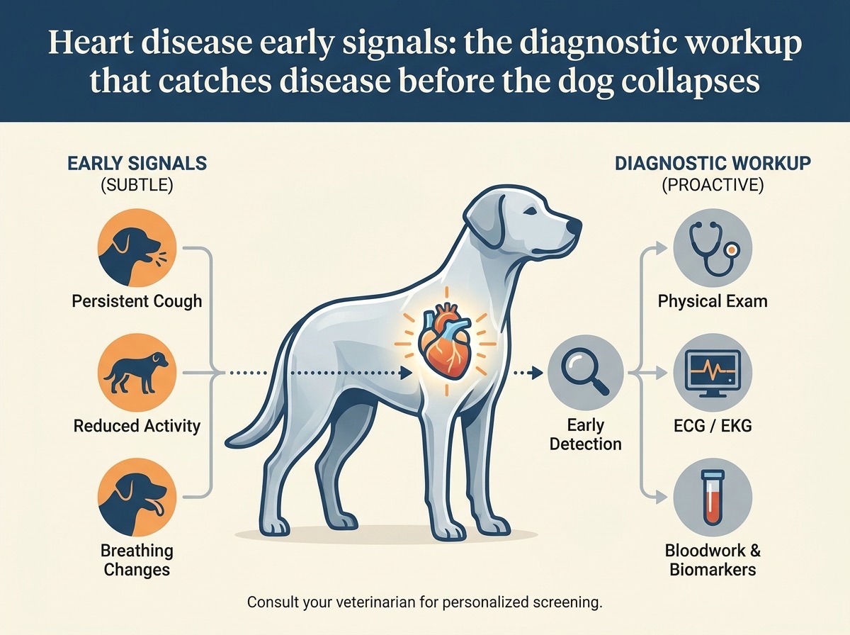
Heart disease early signals: the diagnostic workup that catches disease before the dog collapses
Heart disease in dogs often goes unnoticed until the late stages. The early symptoms are subtle — a slight cough at night, a mild decrease in exercise tolerance, a small weight loss. By the time the dog is actually struggling to breathe, the heart disease is advanced, the cardiac damage is significant, and the prognosis is reduced. The dog that collapses in the yard from heart failure is the dog whose disease was diagnosed too late.
The good news: heart disease in dogs is detectable, treatable, and manageable when caught early. The diagnostic workup is well-established and widely available. The dogs that are screened annually and referred for cardiac workup at the first sign of a problem are the dogs that live 2-4 years longer than the dogs that are diagnosed in crisis.
This is what to watch for, what the diagnostic workup includes, and what the results mean.
The types of canine heart disease
The most common heart diseases in dogs:
Mitral valve disease (MVD)
The most common heart disease in dogs. The mitral valve is the valve between the left atrium and the left ventricle. In MVD, the valve degenerates, becomes thickened and "floppy", and doesn't close properly. Blood leaks backward (regurgitates) from the ventricle into the atrium with each heartbeat. The result:
- Murmur: a "swishing" sound heard on auscultation, caused by the turbulent blood flow
- Heart enlargement: the left atrium and left ventricle enlarge to accommodate the extra blood volume
- Pulmonary congestion: eventually, fluid backs up into the lungs (pulmonary oedema), causing coughing and breathing difficulty
- Heart failure: in late stages, the heart can't pump enough blood to meet the body's needs
MVD is most common in small breeds:
- Cavalier King Charles Spaniel (highest prevalence — nearly all develop MVD by age 10)
- Dachshund
- Miniature Poodle
- Yorkshire Terrier
- Maltese
- Chihuahua
- Cocker Spaniel
MVD is also common in older medium and large breeds, but less predictably than in small breeds.
Dilated cardiomyopathy (DCM)
A disease of the heart muscle. The muscle weakens, the heart chambers enlarge, and the heart's pumping function declines. The result:
- Weak contractions: the heart can't pump effectively
- Chamber enlargement: the heart becomes large and floppy
- Arrhythmias: the heart's electrical system is affected
- Heart failure: progressive decline
DCM is most common in large breeds:
- Doberman Pinscher (highest prevalence — affects a significant proportion)
- Boxer
- Great Dane
- Irish Wolfhound
- German Shepherd
Some breeds have specific genetic mutations associated with DCM. Dobermans, Boxers, and Great Danes have been studied for these mutations.
Other heart diseases
Less common heart diseases include:
- Patent ductus arteriosus (PDA): a congenital defect, usually diagnosed in puppies
- Pulmonic stenosis: a congenital narrowing of the pulmonic valve, often in Bulldogs and Bull Terriers
- Aortic stenosis: a congenital narrowing, often in Golden Retrievers, Boxers
- Heartworm disease: caused by Dirofilaria immitis, transmitted by mosquitoes
- Pericardial effusion: fluid around the heart, often in Golden Retrievers
- Cardiac tumours: usually older dogs, often haemangiosarcoma
The most common diseases in adult dogs are MVD and DCM. The diagnostic workup focuses on these.
The early signals: what to watch for
The early signals of heart disease in dogs are subtle. The owner may notice:
Signal 1: cough at night
A soft, persistent cough at night or early in the morning is one of the earliest signs of MVD. The cough is caused by fluid accumulation in the lungs (pulmonary congestion) or by compression of the main bronchi by the enlarged left atrium.
The owner thinks: "It's just a kennel cough" or "He's been drinking from the water dish too fast". The owner doesn't connect the cough to heart disease.
The vet thinks: a heart murmur, an enlarged left atrium on radiographs, NT-proBNP elevated. The diagnostic workup is initiated.
Signal 2: decreased exercise tolerance
The dog that used to enjoy long walks but now stops after a block. The dog that used to play fetch but now tires quickly. The dog that used to run with the owner but now lags behind.
The owner thinks: "He's just getting older" or "It's the hot weather". The owner doesn't connect the decreased exercise tolerance to heart disease.
The vet thinks: the cardiac output is insufficient to meet the demands of exercise. The diagnostic workup is indicated.
Signal 3: fainting or collapse
The dog that faints during excitement or exercise. The dog that collapses after running. The dog that loses consciousness for a few seconds and then recovers.
The owner thinks: "He just got too excited" or "It was a hot day". The owner doesn't connect the faint to heart disease (or arrhythmias).
The vet thinks: arrhythmia or severe cardiac dysfunction. The diagnostic workup is urgent.
Signal 4: rapid breathing at rest
The dog that is panting at rest, particularly at night. The respiratory rate at rest is normally 15-30 breaths per minute. A sustained rate above 40 in a dog at rest is concerning. Above 60 is an emergency.
The owner can measure the sleeping respiratory rate. This is the single most useful home monitoring tool for dogs with heart disease.
Signal 5: weight loss or muscle wasting
The dog that is losing weight despite eating normally. The dog that has prominent ribs and spine, particularly the "cardiac cachexia" pattern — loss of muscle mass in the back legs, the shoulders, the head.
The owner thinks: "He's getting older and not eating as much". The owner doesn't connect the weight loss to heart disease.
The vet thinks: cardiac cachexia, a wasting syndrome associated with advanced heart disease.
Signal 6: distended abdomen
The dog's belly is larger than usual. The abdomen feels fluid-filled or firm. This is ascites — fluid accumulation in the abdomen.
The owner thinks: "He's gaining weight" or "He's eating too much". The owner doesn't connect the distension to heart disease (typically right-sided heart failure).
The vet thinks: right-sided heart failure. Diagnostic workup indicated.
Signal 7: pale or blue gums
The dog's gums are pale, blue, or mottled. The colour indicates poor perfusion or low oxygen.
The owner thinks: "He just had a dental" or "He's tired". The owner doesn't connect the colour to heart disease.
The vet thinks: severe cardiac or respiratory compromise. Urgent diagnostic workup needed.
The diagnostic workup
The diagnostic workup for suspected heart disease includes several components.
Auscultation (listening with a stethoscope)
The most basic and most important test. The vet listens to the heart and identifies:
- Murmur: a swishing or whooshing sound caused by turbulent blood flow. Murmurs are graded 1-6 by intensity (1 = barely audible, 6 = loud enough to feel as a vibration)
- Arrhythmia: an irregular heart rhythm. The vet notes the rate and the pattern
- Rate: the heart rate. Too fast or too slow are both concerning
- Sounds: extra sounds (gallop rhythms, clicks) may indicate specific conditions
Auscultation is not diagnostic on its own. A murmur indicates the possibility of valve disease but doesn't tell the vet how severe the disease is. Additional tests are needed.
Chest radiographs (X-rays)
The chest X-ray is the most important imaging test for heart disease. It shows:
- Heart size: an enlarged heart (cardiomegaly) indicates significant disease. The vertebral heart score (VHS) is the standard measurement
- Left atrial enlargement: the left atrium enlarges in MVD. The compression of the main bronchi (dorsal displacement) is a sign
- Pulmonary oedema: fluid in the lungs appears as increased density. The pattern helps identify the type (cardiogenic vs non-cardiogenic)
- Pulmonary vessels: enlarged pulmonary veins indicate left-sided heart failure
- Other thoracic structures: the lungs, the diaphragm, the airways are all visible
The chest X-ray is the foundation of the diagnostic workup. It identifies the severity of the disease and the presence of failure.
Echocardiography (ultrasound of the heart)
The gold standard for cardiac diagnosis. The echo provides:
- Chamber size: measurements of the heart chambers, showing enlargement
- Wall thickness: thicker or thinner walls indicate specific conditions
- Valve function: the valves can be visualised directly. Regurgitation can be seen
- Pumping function: the ejection fraction (EF) or fractional shortening (FS) measures the heart's pumping ability
- Doppler blood flow: shows the direction and velocity of blood flow through the heart
Echocardiography requires specialised equipment and a trained operator. Most general practices refer to a cardiologist or a mobile ultrasonographer. The cost is $300-600 for a complete study.
NT-proBNP (cardiac biomarker)
A blood test that measures the level of NT-proBNP, a hormone released by the heart in response to stretch (pressure overload). The result:
- Normal: rules out significant heart disease (high negative predictive value)
- Elevated: indicates heart disease, but doesn't specify the type
- High: indicates significant heart disease and possible heart failure
NT-proBNP is useful for:
- Screening middle-aged dogs for heart disease
- Distinguishing cardiac causes of respiratory signs from respiratory causes
- Monitoring the progression of heart disease
The test is widely available and the cost is reasonable ($50-100). It's a useful first-line screening tool for at-risk breeds.
Electrocardiography (ECG)
Records the electrical activity of the heart. Used to:
- Identify arrhythmias (irregular rhythms)
- Identify chamber enlargement (specific patterns in the ECG)
- Monitor response to antiarrhythmic medications
The ECG is performed at the vet's office or by a cardiologist. A Holter monitor (24-hour ECG) can be used to diagnose intermittent arrhythmias.
Blood pressure measurement
Hypertension is common in dogs with heart disease (and also contributes to kidney disease). The blood pressure is measured similarly to humans:
- Cuff on the limb
- Doppler or oscillometric measurement
- Multiple readings for accuracy
Normal canine blood pressure: systolic 110-140 mmHg. Hypertension: sustained systolic above 160 mmHg.
Bloodwork
Standard bloodwork is performed to assess for:
- Concurrent kidney disease (common with heart disease)
- Liver function
- Electrolyte abnormalities
- Anaemia (worsens cardiac disease)
The bloodwork also provides a baseline before starting cardiac medications.
The staging: how the vet assesses severity
Once the diagnostic workup is complete, the heart disease is staged. The staging system (modified ACVIM consensus):
Stage A: at risk, no disease
- The dog is at risk (breed predisposition, age) but has no identifiable heart disease
- No treatment
- Annual screening
Stage B1: early disease, no symptoms, no significant enlargement
- Murmur present (in MVD)
- Heart not significantly enlarged on X-ray or echo
- No symptoms
- No treatment indicated
- Recheck every 6-12 months
Stage B2: moderate disease, no symptoms, significant enlargement
- Murmur present
- Heart significantly enlarged on X-ray (VHS above breed-specific threshold) or echo
- No clinical symptoms
- Treatment indicated: pimobendan (Vetmedin) to slow progression
- ACE inhibitor (enalapril, benazepril) is sometimes started at this stage
Stage C: heart failure, current or recent
- Symptoms present or recently resolved
- Pulmonary oedema, cough, exercise intolerance
- Treatment: combination of pimobendan, ACE inhibitor, diuretic (furosemide), and other medications as needed
- Careful monitoring
Stage D: refractory heart failure
- Heart failure that is not responding to standard treatment
- Severe symptoms despite medications
- Treatment: advanced medications (pimobendan + ACE inhibitor + spironolactone + additional diuretics), possible hospitalisation
- Quality of life is the focus
The stage determines the treatment. The treatment changes as the disease progresses.
The treatment: what's available
The treatment options depend on the stage and the type of disease.
Pimobendan (Vetmedin)
The most important medication for MVD and DCM. Pimobendan:
- Increases the heart's contractility (positive inotrope)
- Dilates the blood vessels (vasodilator)
- Reduces the heart's workload
In MVD: pimobendan started at stage B2 (before symptoms) extends the time to heart failure or death by approximately 15 months (the EPIC study, 2016).
In DCM: pimobendan improves survival in dogs with congestive heart failure.
ACE inhibitors (enalapril, benazepril, lisinopril)
- Dilate blood vessels, reducing the heart's workload
- Reduce fluid retention indirectly
- Protect the kidneys (important in dogs with concurrent kidney disease)
ACE inhibitors are commonly used at stage C and D. They are sometimes started at B2.
Diuretics (furosemide, torsemide)
- Remove excess fluid from the lungs and abdomen
- Critical for treating pulmonary oedema (the life-threatening complication of heart failure)
Furosemide is started when fluid accumulates (stage C). The dose is adjusted based on the severity of the congestion. The owner monitors the dog's respiratory rate at home.
Spironolactone
- Potassium-sparing diuretic
- Mild cardiac protective effects
- Used in combination with furosemide at stage C and D
Other medications
- Amlodipine: calcium channel blocker, used for hypertension
- Sildenafil: PDE5 inhibitor, used for pulmonary hypertension
- Antiarrhythmic drugs: for specific arrhythmias (sotalol for Boxer cardiomyopathy, etc.)
- Antiplatelet drugs: clopidogrel, for dogs with cardiac disease at risk of thromboembolism
Diet
- Sodium restriction for dogs in heart failure
- Cardiac prescription diets (Hill's h/d, Royal Canin Cardiac)
- Omega-3 supplementation for cardiac cachexia
The home monitoring: the owner's role
The owner can monitor the dog at home to detect early signs of failure or progression:
Sleeping respiratory rate
The most useful home monitoring tool. The owner counts the breathing rate while the dog is sleeping (or calm and relaxed). The normal rate is 15-30 breaths per minute. A sustained rate above 30-35 is concerning. Above 40 indicates possible heart failure.
The owner counts for 15-30 seconds and multiplies. The count is logged. The vet reviews the log. Trends are more useful than single readings.
Cough frequency
The owner notes the frequency and severity of any coughing. A sudden increase in coughing is a sign of worsening disease or pulmonary congestion.
Exercise tolerance
The owner notes the dog's ability to exercise. A decrease in exercise tolerance is a sign of worsening disease.
Weight
The owner weighs the dog regularly (weekly). A rapid weight gain may indicate fluid retention. A weight loss may indicate cardiac cachexia.
Appetite and behaviour
The owner notes changes in appetite, energy, and behaviour. A decrease in appetite may indicate nausea (a side effect of medications or a sign of progressing disease).
The screening recommendations
For at-risk breeds:
- Cavalier King Charles Spaniel: annual cardiac auscultation starting at age 2-3. Echocardiography if a murmur is detected. This breed has the highest prevalence of MVD
- Doberman Pinscher: annual cardiac screening starting at age 2-3, including auscultation, echocardiography, and Holter monitor (24-hour ECG). This breed has the highest prevalence of DCM
- Boxer: annual screening including Holter monitor. Boxers can have arrhythmogenic cardiomyopathy
- Other small breeds (MVD): annual auscultation starting at age 5-7
- Other large breeds (DCM): annual screening starting at age 3-5
For non-at-risk breeds, the cardiac screening is part of the annual senior wellness screen (recommended at age 7+).
The "what to do if heart disease is suspected" checklist
The owner who notices any of the early signals:
- Schedule a vet visit within 1-2 weeks for auscultation
- Note the symptoms (cough frequency, exercise tolerance, sleeping respiratory rate)
- Bring records of any prior heart evaluations
- Ask for a cardiac workup if a murmur is detected
- See a cardiologist if the disease is moderate or advanced
- Start medications as directed
- Monitor at home (sleeping respiratory rate, weight, exercise tolerance)
- Recheck regularly as directed
The "the dog collapsed, what now" emergency
A dog that has collapsed from heart failure is an emergency. The action:
- Stay calm: the dog is in distress
- Keep the dog calm: minimise stress and movement
- Transport to the nearest emergency clinic immediately: call ahead so the team is ready
- Bring the dog's medications: pimobendan, furosemide, etc. The emergency vet will need to know what the dog is taking
- The emergency vet will perform chest X-rays, bloodwork, oxygen therapy, IV medications (furosemide, pimobendan), and possibly sedation
The acute management of heart failure is well-established. The dog can be stabilised in most cases. The underlying disease is the issue, and the dog may recover with treatment.
The bottom line
Heart disease in dogs is common, manageable, and detectable when caught early. The diagnostic workup — auscultation, chest X-rays, echocardiography, NT-proBNP, bloodwork — is widely available and provides a comprehensive assessment. The staging system guides treatment. The early stages are managed with monitoring and minimal medication. The late stages are managed with combination therapy. The dogs that are screened annually and referred early live 2-4 years longer than the dogs that are diagnosed in crisis. The owner who understands the early signals and pursues the diagnostic workup is the owner who gives the dog the longest life. The vet is the partner. The cardiologist is the resource. The home monitoring is the daily work. The disease is the enemy. The treatment is the weapon. The dog is worth it.
---
About the author: [James Morrison](../James%20Morrison.md) holds a BVSc from the University of Liverpool and has 22 years of small animal general practice, with a focus on preventive medicine and multi-pet household dynamics.