

Foreign body ingestion in dogs and cats: when to X-ray, when to scope, when to wait
Foreign body ingestion is the second most common emergency I see in my practice (after hit-by-car). The decision tree for what to do next is one of the most useful things a pet owner can understand, because the wrong call at home — either rushing in for an emergency surgery that wasn't needed, or waiting at home when surgery was urgent — has consequences in both directions.
This is the framework I walk owners through when they call me saying "we think he ate something".
The three immediate questions
When a pet owner calls me about a possible foreign body, the first three questions are always the same:
1. What was swallowed?
Knowing the object determines the urgency:
- Sharp objects (needles, pins, bone fragments, glass): urgent. Risk of perforation
- Long linear objects (string, yarn, ribbon, tinsel): urgent in cats especially. Risk of linear foreign body with plication (the intestine gathers accordion-style along the string)
- Toxins (batteries, lead, zinc-containing objects, medications): urgent. Toxins absorb directly
- Soft objects (sock, underwear, plush toy fragment, fabric): variable. Many pass on their own
- Hard non-toxic objects (coin, marble, small stone, plastic Lego): variable
- Food objects (corn cob, peach pit, bone fragment): variable, but bones often cause perforation
2. How big is it relative to the pet?
A 5 cm sock in a 40 kg dog is very different from a 5 cm sock in a 3 kg cat. The smaller the pet, the more likely the object will obstruct.
A rough size threshold:
- <3 cm in a cat: high obstruction risk
- 3–5 cm in a cat or small dog: moderate obstruction risk
- >5 cm in a dog >15 kg: usually passes
3. How long ago was it swallowed?
- <2 hours: object may still be in the stomach. Endoscopic retrieval is feasible.
- 2–12 hours: object may be in the small intestine. Endoscopy is no longer possible (the scope only reaches the stomach and duodenum). Monitoring or surgery becomes the path.
- >12 hours: object has likely moved beyond the stomach. Monitoring or surgery depending on signs.
- >24 hours without passage: increased risk of obstruction, perforation, or both.
These three answers give me about 70% of the information I need to advise the owner on next steps.
The X-ray: when, why, and what it shows
Plain radiographs (X-rays) are the first-line imaging for suspected foreign bodies. They show:
- Radiopaque objects clearly: metal, bone, stone, some plastics (with strong contrast), coins
- Obstruction signs: dilated bowel loops, gas patterns, faecal stasis
- Linear foreign body pattern: plicated bowel, gathering of intestinal loops
- Pneumoperitoneum: free air in the abdomen, indicating perforation (emergency)
What X-rays do NOT show:
- Radiolucent objects: most cloth, fabric, wood, rubber, some plastic, fish bones
- Subtle obstruction signs in early disease
The radiopaque vs radiolucent distinction is critical:
| Easily seen on X-ray | Often missed on X-ray |
|---|---|
| Metal (coins, batteries, hooks, pins) | Cloth (sock, underwear, plush fabric) |
| Bone fragments | Wood splinters |
| Stones | Rubber balls |
| Glass (sometimes) | Plastic toys (most types) |
| Dense plastics | String, yarn, ribbon |
For radiolucent objects, contrast X-rays or ultrasound are needed.
Contrast X-rays: the bariums and Gastrografin
If the object is radiolucent and the pet is stable, a contrast study can be useful:
- Barium sulphate: administered orally as a paste or liquid. Follow-up X-rays at 30 min, 1 hour, 2 hours, 4 hours, 6 hours, 12 hours
- Gastrografin (diatrizoate): water-soluble contrast, useful when perforation is suspected (barium in the abdomen is bad if perforation present)
The contrast study shows where the GI tract is patent (contrast flows through) and where it is obstructed (contrast pools). It takes hours of waiting and serial X-rays.
In modern practice, contrast studies have been largely replaced by ultrasound and endoscopy. The contrast study is still useful when ultrasound is unavailable or inconclusive.
Ultrasound: the underused workhorse
Abdominal ultrasound is often more useful than X-ray for foreign body cases:
- Detects radiolucent objects: cloth, wood, plastic often visible on ultrasound even when missed on X-ray
- Detects obstruction signs earlier: dilated bowel loops, fluid-filled intestines, decreased peristalsis
- Identifies perforation signs: free abdominal fluid, bowel wall thickening, loss of wall layering
- Can guide endoscopy or surgery: localises the object
The limitation: ultrasound is operator-dependent. A skilled sonographer can identify most foreign bodies; a less experienced one may miss subtle findings.
In my practice, the diagnostic path for suspected foreign body is:
- Plain X-rays (2 views: lateral and VD): first-line, often diagnostic for radiopaque objects
- Abdominal ultrasound: if X-rays are negative or equivocal and the pet is symptomatic
- CT scan: if both X-rays and ultrasound are inconclusive and clinical suspicion remains high
When to wait, when to act
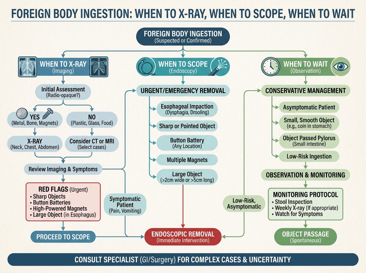
This is the most important question for owners. The decision tree:
Asymptomatic pet, recent ingestion (<2 hours), small object
If the pet is eating normally, not vomiting, and behaving normally, observation is often appropriate. Most small objects pass within 24–48 hours. The owner should:
- Feed bulky food (canned pumpkin, bread, normal meals) to help push the object through
- Check stool for passage (wear gloves, use a stick)
- Bring the pet back immediately if vomiting, lethargy, or appetite loss develops
Asymptomatic pet, but sharp or linear object
Sharp or linear objects warrant more aggressive action even without symptoms:
- Sharp object (needle, pin, glass): X-ray to confirm location, monitor closely, endoscopy if in the stomach
- Linear object in a cat (string, yarn): this is a surgical urgency in many cases. Linear foreign bodies plicate the bowel and cause necrosis quickly. ER visit recommended
Symptomatic pet (vomiting, anorexia, lethargy)
Symptomatic foreign body is a medical urgency:
- X-ray + ultrasound to identify the object and location
- Bloodwork: elevated PCV (dehydration), elevated lactate (tissue hypoperfusion or necrosis), electrolyte disturbances
- IV fluids, anti-nausea medication, pain management while diagnostics are in progress
- Surgery or endoscopy based on findings
Toxic object (battery, lead, medication, zinc)
Toxic objects are emergencies:
- Button batteries: caustic, can cause perforation within hours. Endoscopy or surgery immediately
- Lead objects: lead toxicity develops over days. Remove if possible, monitor blood lead levels
- Medications (even human OTC): toxic dose depends on the medication and the pet's weight. Call poison control
- Zinc objects (pennies minted after 1982, zinc-containing objects): zinc toxicity causes severe anaemia. Remove if possible
Endoscopy: when it works
Endoscopic foreign body retrieval works only if:
- The object is in the stomach or duodenum (the scope reaches this far)
- The object is small enough to pass through the oesophagus on the way out (or can be grasped and pulled back)
- The object is not sharp in a way that will lacerate the oesophagus on retrieval
- The pet is stable enough for anaesthesia
Endoscopy advantages over surgery:
- No abdominal incision
- Faster recovery (often same-day discharge)
- Lower cost (typically 50–70% of surgical cost)
- Lower complication rate
Endoscopy disadvantages:
- Not all objects are reachable
- Some objects are too large or too sharp for endoscopic retrieval
- Anaesthesia is still required
- Equipment and skill not available everywhere
When surgery is necessary
Surgery is indicated when:
- The object is in the small intestine or colon (out of endoscopic reach)
- The pet has signs of obstruction with object confirmed on imaging
- There are signs of perforation (pneumoperitoneum, septic peritonitis)
- The object is sharp and likely to cause perforation
- 24+ hours have passed without passage in a symptomatic pet
- Endoscopy is unsuccessful
The most common surgical procedures:
- Gastrotomy: opening the stomach to retrieve a gastric foreign body
- Enterotomy: opening the intestine to retrieve a small intestinal foreign body
- Resection and anastomosis: removing a section of intestine that has died from obstruction, and rejoining the healthy ends
- Linear foreign body surgery: multiple enterotomies to remove a long linear object without cutting the bowel
The surgical complication rate has dropped dramatically in the last 20 years. Survival rates:
- Simple gastrotomy/enterotomy: >95% with experienced surgeon
- Resection and anastomosis: 80–90%
- Septic peritonitis from perforation: 50–70% (depends on duration)
Linear foreign bodies in cats: a special warning
Cats and string are a dangerous combination. Cats (especially young cats) love to play with string, tinsel, ribbon, thread, dental floss, and similar items. They often swallow them.
The danger is the linear foreign body with plication: the string gets anchored at one end (often under the tongue, around the base of the tongue, or at the pylorus) and the intestine gathers accordion-style along it. This causes:
- Intestinal plication: the bowel folds around the string
- Mesenteric tears: as the intestine gathers, the mesentery can tear
- Perforation: the string slowly saws through the intestinal wall
- Necrosis: the affected bowel segment dies
Signs in cats:
- Vomiting (often intermittent, may produce food, bile, or foam)
- Anorexia: the cat stops eating
- Lethargy: hiding, less interactive
- String visible under the tongue or around the anus (a classic finding, always check)
- Pain on abdominal palpation: a tense, painful abdomen
If you suspect your cat has swallowed string, ribbon, tinsel, or thread:
- Check under the tongue: gently lift the cat's upper lip and look at the base of the tongue. If string is visible, do NOT pull it (cutting is safer than pulling)
- Seek emergency veterinary care within hours: linear foreign bodies can become surgical within 12–24 hours
- Do not wait for the cat to "pass it" — many don't
Monitoring after discharge
For pets that are sent home without intervention, the monitoring protocol:
- Stool checks: wear gloves, use a stick, check stool for the object. Most small objects pass within 24–72 hours
- Appetite monitoring: any drop in appetite for >12 hours warrants a vet visit
- Vomiting: any vomiting (not just retching) is a red flag
- Lethargy: any change in energy level is a red flag
- Abdominal pain: pet flinches when belly is touched, adopts a "prayer position" (front down, hindquarters up) to relieve abdominal discomfort
The rule: if the pet is not back to normal within 24 hours of ingestion, see a vet.
Prevention
The cases that don't need to happen:
- Keep small objects out of reach: socks, underwear, hair ties, toys with small parts
- Pet-proof the laundry basket: dogs that eat socks usually start with the laundry basket
- Choose appropriately-sized toys: toys that fit entirely in the mouth can be swallowed. The rule: if the toy cannot fit past the dog's lower jaw, it is too small
- Cats and string: keep sewing supplies, tinsel, ribbon, dental floss, and yarn out of cat reach. The "string toy" sold for cats is itself a risk — supervise play and put away when not actively playing
- X-ray toys: avoid toys with small magnets. Multiple magnets in the GI tract attract each other through the bowel wall and cause perforation
- Cooked bones: never give cooked bones. They splinter. Raw bones are larger and softer but still risk
The bottom line
Foreign body ingestion is a common, usually solvable emergency. The decision tree is: know the object, know the size, know the timing. Asymptomatic recent small ingestion: monitor. Asymptomatic sharp or linear: act fast. Symptomatic: ER visit with X-ray and ultrasound. Toxic object: emergency endoscopy or surgery. Linear foreign body in cat: surgical urgency. The cases that do well are the ones where the owner recognised the risk early and the pet was assessed within hours, not days. The cases that don't are the ones where everyone waited for the pet to "pass it" while the bowel was dying.
---
About the author: [Dr. Sofia Martinez](../Sofia%20Martinez.md), DVM, CertECC, is a UCM-trained veterinarian with 13 years of practice and former head of emergency surgery at Hospital Veterinari 24h in Barcelona.