

Malassezia Dermatitis in Dogs and Cats: How to Spot It at Home Before It Spreads
If your dog has started smelling like Fritos, or your cat is scratching under the arms until the skin turns black, you may be looking at Malassezia — a yeast that lives on virtually every pet's skin, and turns pathological when the surface environment tips in its favor. As a veterinary dermatologist, I see this condition several times a week, and in most cases the owner noticed something was wrong two to four weeks before they brought the pet in. This guide is designed to help you recognize those early signals, run a defensible at-home response, and — most importantly — know exactly when to stop home treatment and escalate.
What Malassezia Actually Is
Malassezia pachydermatis is a lipophilic (fat-loving) yeast that colonizes the skin, ear canals, anal sacs, interdigital spaces, and ventral neck fold of most healthy dogs and cats. It does not "infect from outside" — it overgrows from the resident population when the local microenvironment changes. The three triggers I see most often in clinic are:
- Underlying allergy (atopic dermatitis, food allergy, flea allergy) — the most common driver by a wide margin.
- Moisture and skin fold occlusion — Bulldogs, Shar-Peis, Basset Hounds, Pugs, and overweight cats with redundant ventral skin.
- Recent antibiotic or steroid use — disrupts the bacterial–yeast balance and removes competing flora.
Cats also get Malassezia, but the presentation is more subtle and is almost always secondary to a primary disease such as feline atopic syndrome, food allergy, or an internal disorder (especially upper respiratory disease with facial rubbing).
The Four Visual Clues You Can Confirm at Home
You do not need a microscope to triage. Malassezia has a recognizable signature:
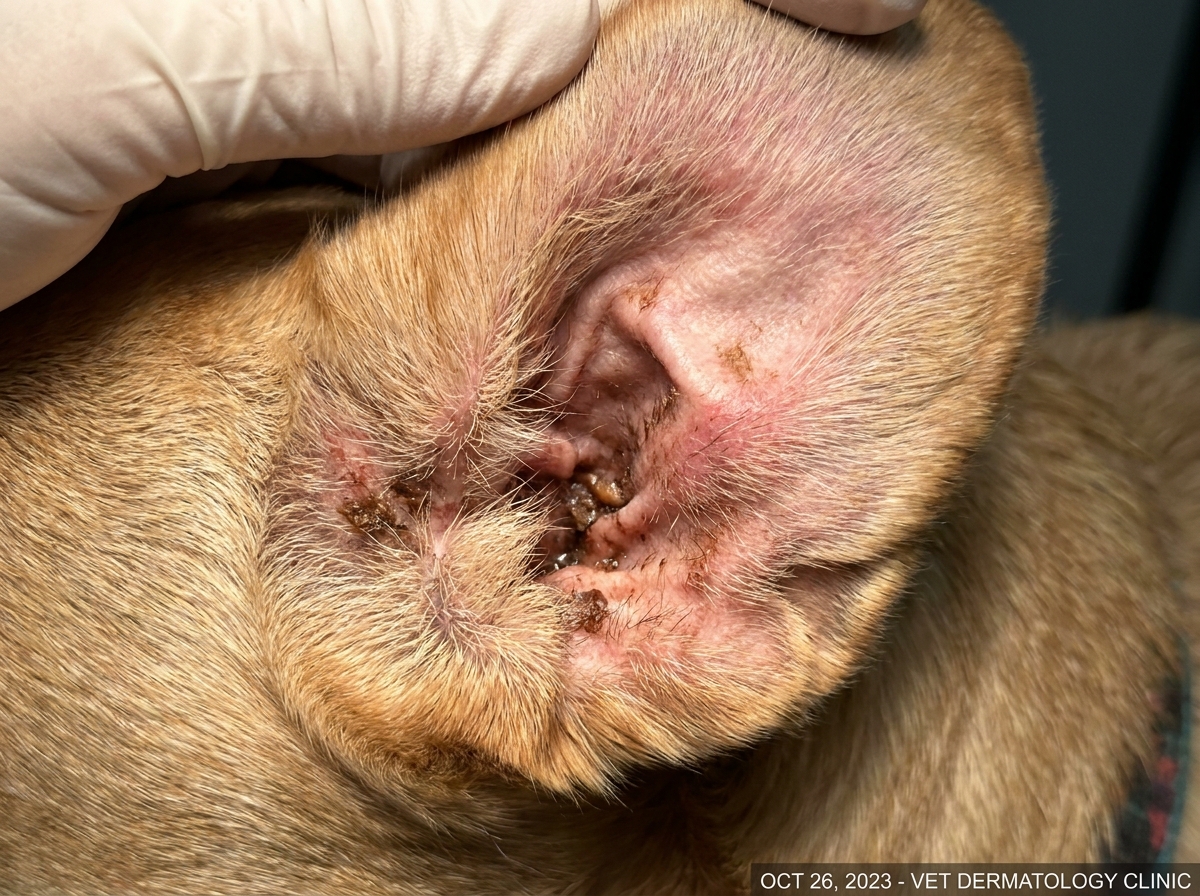
- Color: a greasy, dark brown to rust-colored exudate in the ear canals, under the armpits, in the groin, between the toes, and in the skin folds of the face and neck. In cats, the chin and ventral abdomen often turn black ("hyperpigmentation") rather than red.
- Texture: waxy, oily, sometimes described as "damp cornflakes." It does not flake the way dry seborrhea does.
- Odor: a sweet, musty, slightly rancid smell — the "Frito feet" reference is real and is almost pathognomonic in dogs.
- Itch level: high. If your pet is awake scratching the same spot for more than 30 minutes a day, or you see saliva staining on the front paws, count this as a positive signal.
A simple at-home test: press a clear piece of tape against the affected area, lift it, and lay it on a white piece of paper. Thick brownish-yellow residue that transfers in clumps is suggestive. This is not a substitute for cytology, but it is a useful way to decide whether the trip to the vet is urgent or can wait for the next available appointment.
Triage: Green, Yellow, Red
Before reaching for any medication, run this triage:
- Green (home care for 5–7 days is reasonable): mild odor, redness confined to one area (one ear, one paw, the groin), no broken skin, eating and behaving normally.
- Yellow (call the clinic within 24–48 hours): spreading to two or more body regions, more than a 50% increase in scratching, recurrent head-shaking, or any odor change combined with hair loss.
- Red (same-day visit or ER): open sores, bleeding, hot or painful skin, fever, lethargy, refusal to eat, or a pet in obvious distress. Malassezia is rarely a true emergency, but the secondary bacterial infection that often rides with it (especially in dogs with deep folds) can be.
What to Try at Home — and What to Skip
If you are in the green zone, a two-step plan is reasonable:
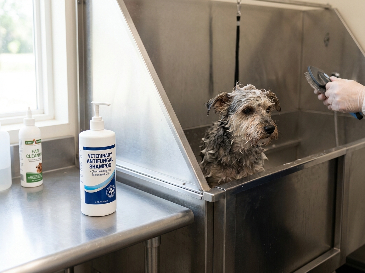
- Antifungal shampoo bath every 2–3 days for 3 weeks, then weekly for 3 more weeks. The two products with the strongest evidence are:
- 2% chlorhexidine + 2% miconazole (commonly sold as Malaseb or generic equivalents). Lather, leave the suds on the skin for 10 minutes, then rinse. The 10-minute contact time is non-negotiable.
- 3–4% chlorhexidine alone as a second-line option if miconazole is unavailable.
- Antifungal ear cleaner for any pet with concurrent ear odor. Look for products containing chlorhexidine 0.15% + miconazole 0.15% or ketoconazole 1%. Use daily for 7 days, then every other day for 3 weeks.
Do not use human over-the-counter antifungals like clotrimazole cream (too weak against Malassezia), tea tree oil (toxic to cats at therapeutic doses), or hydrogen peroxide (damages healing skin). Do not use topical antibiotic-only ointments — they do not touch yeast and may worsen the imbalance.
When the Clinic Has to Take Over: Named Medications and Real Doses
If the green-zone plan does not produce a 30–50% improvement in odor and itch by day 7, your pet needs cytology and probably systemic therapy. The medications I reach for most often, with the doses I actually prescribe, are:
| Drug | Indication | Dose (dogs) | Dose (cats) | Key note |
|---|---|---|---|---|
| Ketoconazole | Generalized Malassezia | 5–10 mg/kg PO q12h with food | 5 mg/kg PO q12h with food | Do not use in cats with liver disease; always give with a meal for absorption. |
| Itraconazole | Ketoconazole-intolerant patients | 5 mg/kg PO q24h with food | 5 mg/kg PO q24h with food | Pulse therapy (1 week on / 1 week off) often works after induction. |
| Terbinafine | Refractory or relapsing cases | 30 mg/kg PO q24h with food | 20–30 mg/kg PO q24h | Monitor liver enzymes after 4 weeks. |
| Oclacitinib (Apoquel) | When itch is allergy-driven and Malassezia is secondary | 0.4–0.6 mg/kg PO q12h for up to 14 days, then q24h | Not approved in cats | Controls pruritus so the skin can actually heal. |
| Cytopoint (lokivetmab) | Dogs with atopic flare and concurrent yeast | 2 mg/kg SC, repeated every 4–8 weeks as needed | Not approved in cats | A good alternative when oral drugs are hard to give. |
The hard rule: never combine ketoconazole with cyclosporine (Atopica) without an explicit drug-interaction check. Ketoconazole blocks CYP3A and will roughly triple cyclosporine blood levels, which can cause kidney injury.
When to Escalate — The Exact Thresholds
Call your vet the same day if any of the following appear during home treatment:
- The skin becomes hot, painful, or has a foul pus-like smell (suspect secondary bacterial pyoderma).
- Itch has not improved at all by day 5, or has worsened by day 3.
- The pet develops vomiting, inappetence, or yellowing of the gums or sclera after starting any oral antifungal.
- The pet is a cat with any blackening of the chin or ventral abdomen — feline Malassezia almost always reflects a primary disease (allergy, viral, endocrine) that needs diagnostic workup, not just topical therapy.
- Recurrence happens more than three times a year — this is a signal to look for an underlying allergy, not to keep shampooing.
The Bottom Line
Malassezia is a manageable condition, but it is almost always a messenger. Your job at home is to recognize the smell, the brown wax, and the itch early, run a measured 7-day shampoo-and-cleaner protocol if the case is mild, and stop home treatment the moment the green zone tips toward yellow. Treating the yeast without diagnosing the underlying allergy is the single most common reason I see relapses in clinic. If the smell comes back within a month, book a follow-up specifically to discuss allergy workup — elimination diet trial, intradermal skin testing, or serum IgE panel — not just another round of shampoo.
---
About the author
Hannah Kim, DVM, DACVD, is a board-certified veterinary dermatologist. She has treated over 4,000 cases of Malassezia dermatitis across dogs, cats, and rabbits. This article reflects her clinical practice as of July 2026 and is intended for general educational purposes — it does not replace an in-person examination.
References (selected)
- Bond R, et al. Malassezia dermatitis in dogs: a review. Vet Dermatol. 2022;33(4):289-302.
- Morris DO, et al. Treatment of Malassezia dermatitis in dogs: a randomized clinical trial. Vet Dermatol. 2021;32(2):165-e34.
- Olivry T, DeBoer DJ, Favrot C, et al. Treatment of canine atopic dermatitis: 2015 updated guidelines from the International Committee on Allergic Diseases of Animals (ICADA). BMC Vet Res. 2015;11:210.